AI Facial Asymmetry Analysis

Ultrasound first,
then the plan.

If one side of your jaw looks bigger, is it muscle or bone?

We check by ultrasound first. We don't treat what we haven't seen.

Book a Scan & Consult

A doctor measures in person. About 5 minutes.

Sample: left and right measured separatelyExample values

Masseter thicknessExample image · made from the doctor's own ultrasound, with only the right muscle layer enlarged

ExampleExample. Right masseter ultrasound, 14.5mm from fascia to jawbone line
Right · Grade 414.5mm
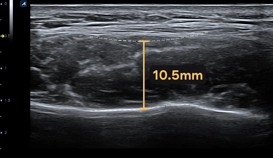
ExampleExample. Left masseter ultrasound, 10.5mm from fascia to jawbone line
Left · Grade 310.5mm

The right side is 4.0mm thicker and one grade higher

So the two sides don't get equal amounts. It's split by the thickness ratio.

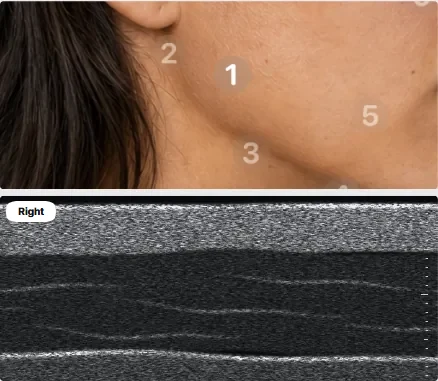
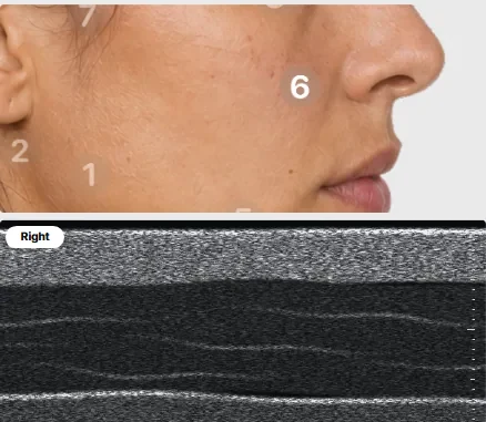
Photo and ultrasound image of the area beside the smile line (nasolabial fold)
Fat beside the smile lineThe fat layer running from beside the nose to the corner of the mouth
Right8.5mm
Left6.8mm

1.7mm apart, over 30% of the 3mm average range width. So the report adds an asymmetry graphic.

See the full 10-area table

Does AI choose the treatment?

No. We split the work into three roles.

The doctor's role

  • Confirms the thickness valuesThe values that go into the ultrasound report
  • Also sets the midline of the 3D faceFrom between the eyes to between the nostrils · Meta-Vu
  • Treatment is decided together, in consultationAI doesn't pick treatments

AI's role

  • Aligns before/after photos to the same position and sizeFinds facial points to match them · Mark-Vu
  • Checks that the 3D scan came out wellTells us if a retake is needed · Meta-Vu
  • Sorts ultrasound images by areaUltrasound report

Doctor + AI

The two combined

  • Asymmetry graphics are added by ruleWhen the left-right difference exceeds 30% of the average range width
  • Suggested treatments also come from rulesRules the doctor set in advance
  • The doctor reviews what AI findsConfirmation comes after that

We aim for the approach that fits you.

How big a difference counts as different?

Each area has its own threshold: 30% of that area's average range width. Only areas past that line get an asymmetry graphic.

Smile line 0.9mm · Deep cheek 1.5mm · Below cheekbone 0.6mm

Left-right bars from a sample report. Left 3mm, right 6mm
Sample report example · a different sample from the table below

Areas past the threshold get left-right bars like these.

Left-right volume comparison from a sample report. Left 47%, right 53%
Sample report example

A left-right ratio combining fat thickness with muscle and salivary gland grades. In consultation, it helps decide how much to address on each side.

Sample table: 10 facial areasExample values

AreaRightLeftDifference
1MasseterMuscle · grade4Grade3Grade1Grade
2ParotidSalivary gland · grade3Grade2Grade1Grade
3Under-jaw glandSalivary gland · grade3Grade2Grade1Grade
4Under-chin fatFat · one central point14.0mm–
5Deep cheek fatFat12.4mm11.2mm1.2mm
6Smile-line fatFat8.5mm6.8mm1.7mm
7Below-cheekbone fatFat4.6mm4.1mm0.5mm
8Under-eyeDermis thickness1.32mm1.28mm0.04mm
9EyelidDermis thickness0.82mm0.54mm0.28mm
10ForeheadDermis thickness1.07mm0.98mm0.09mm

Row 6 shows a left-right difference over 30% of that area's average range width, so the report adds an asymmetry graphic. All values are for illustration, not real patient data.

We look at the dermis tooExample values

Close-up of the dermis on an ultrasound image. The dermis between the upper and lower red lines is colored in 5 shades of green
A sample where the dermis on an ultrasound image is colored in five shades of green by brightness. It's a synthetic image, not a real patient's.
Forehead dermis thickness Measured between the upper and lower red lines. Use the 1mm scale in the image to judge size.
Right1.07mm
Left0.98mm

Dermal density

Right46.1pts
Left46.2pts

Dark cellsBright cells

Dermal density scores, from 0 to 100, how many bright cells show in the dermis on ultrasound. More bright cells, higher score. But scar tissue looks bright too, so a high score doesn't mean good skin, or a low one bad skin. Each age group has its own expected range.

Why is one side wider?

Muscle, fat, bone. We narrow it down in that order. The first two are measured and compared. If both are similar, then we look at bone.

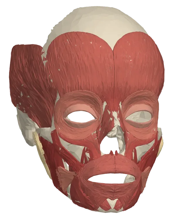
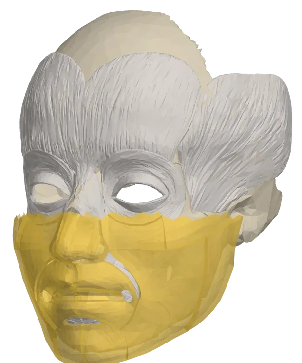
Standard anatomical model with only the masseter shaded Standard anatomical model with lower-face fat layers shaded yellow Standard anatomical model with the lower jaw and cheekbones shaded
A standard anatomical model for illustration, not any individual's face. Adapted from Z-Anatomy (CC BY-SA 4.0), BodyParts3D © DBCLS (CC BY 4.0)
  1. Three layers · standard modelWe check three layers in turn

    Steps 1 to 3 each check one layer. Two layers are measured, and the third is judged from those results.

  2. 1Muscle · masseter · 5-level gradeIs one masseter bigger?

    The jaw muscle that firms up when you clench your back teeth. Each side is measured and graded 1 to 5.

  3. 2Fat · 4 areas · mmIs the fat thicker on one side?

    We record each side's thickness in mm, area by area. If the difference crosses the threshold, it's flagged in the report.

  4. 3Bone · not measured · judged from 1 and 2If both are similar but the jaw sticks out

    If muscle and fat are similar on both sides but one side of the jaw projects more, we consider a bone difference. Bone itself isn't measured by ultrasound.

  5. 4Approach · consultation · with the doctorWe talk through balancing both sides

    Reshaping bone isn't an easy choice. So we start by talking about reducing a little fat to even things out.

What brings you in?

My two sides look different

Left-right ultrasound report · Alpinion E-CUBE 8

We measure 10 areas, from the masseter to the forehead. The report comes in 6 languages, delivered by QR code or link.

More on the ultrasound report
Masseter section of a sample report

My before/after photos never line up

Photo alignment · Mark-Vu

AI finds facial points and matches the position and size of before/after photos, so they sit in the same place side by side.

More on photo alignment
Example Mark-Vu front photo with 8 facial reference points marked. Eyes pixelated
Photos of Wi Young-jin, Chief Director, Myeongdong, used with consent. Eyes are pixelated. White circles = 8 alignment points. When AI aligned the 1st and 16th photo sessions, the average point offset between the two photos dropped from 11.8px to 8.5px. This is photo alignment accuracy, not a change in the face (720px-wide photo).

Can we start with my midline?

3D scan · Meta-Vu

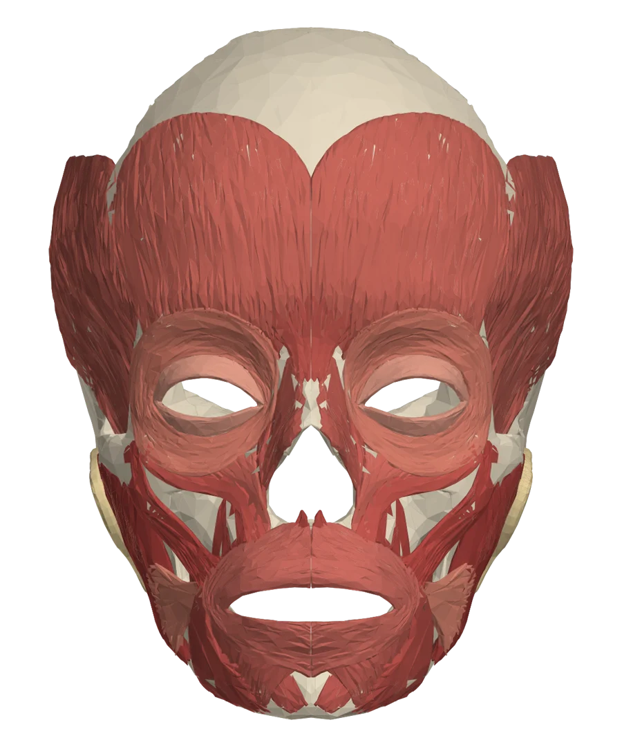
The 3D face is set upright on the midline the doctor defines. The overlay is a standard anatomical model, and thickness is shown roughly in place.

More on the 3D scan
3D standard anatomical model of facial muscles and bones, front view
Front view of muscles, bones, and salivary glands drawn from a generic 3D anatomical model, not a specific person's face. Adapted from Z-Anatomy (CC BY-SA 4.0), BodyParts3D © DBCLS (CC BY 4.0)

Before you're measured

Asymmetry we look at
We measure left-right differences in muscle, salivary glands, and fat under the skin. Bone isn't measured by ultrasound. If muscle and fat are similar and a difference remains, that's when we look at bone and talk it through.
Average range
The average range is a reference value set in the report, and we're checking it against measurements from our Hongdae branch. Below the cheekbone, measured values clustered on the thicker side of the range. Please treat it as a reference line only.
Where suggestions come from
Suggested treatments come from rules the doctor set. Actual treatment is decided in consultation.
Photo processing
Before/after photo alignment is processed on the clinic's server.

Beautystone Clinic · 45 Yanghwa-ro, Mapo-gu, Seoul, Mecenatpolis Mall (Units 133–135, 215–218) · 02-3144-0107 · Measurement and treatment results vary by person, and treatment may cause side effects.

Let's look at the numbers

At our Hongdae branch, we measured 59 times from August 26 to October 31, 2026. In 38 (64%), no area crossed the threshold. In 21 (36%), at least one did. There's no telling which group you'll be in until you're measured.

These are two months of records from one branch, so we don't assume they apply elsewhere as is. You can book just the measurement, and simply hear your results without choosing any treatment.

Book a Scan & Consult
Beautystone

1:1 Custom Approach

1:1 Consultation Whatsapp

🌸 Beautystone Clinic attends the Meditox Bangkok Cadaver Workshop 🌸

1:1 Custom Approach